Alternatives to DEXA Scan Body Composition Analysis
The interest in non-invasive methods of body composition assessment is on the rise in health care, especially because of its association with clinical outcomes.
Technology has revolutionized our understanding of body composition abnormalities, clinical prognosis, and disease follow-up, but their use on non-medical settings are still limited, and they tend to be costly. It is therefore extremely important to understand the advantages and disadvantages of each technique in specific situations before deciding on a body composition assessment technique.
Up to recently, magnetic resonance imaging (MRI) and computed tomography (CT) are considered the gold standard techniques for the assessment of muscle mass/muscle quantity. The reason is that both techniques are cross-sectional and allow for precise evaluation of compartmental body composition at different regions of the body (skin, muscle, and body fat tissues). However, Dual-energy X-ray absorptiometry (DEXA) scan has emerged as the preferred method for body composition analysis thanks to its high accuracy, safety, and ease of use.
In this article you’ll learn about the three most utilized body composition assessment techniques in order to better understand the option that best corresponds to your needs.
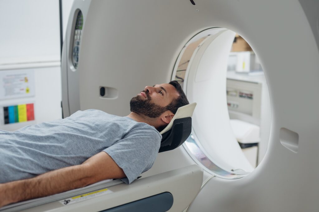
CT Scan
A CT (Computed Tomography) scan is an imaging test that helps healthcare providers detect diseases and injuries. It uses a series of X-rays and a computer to create detailed images of your bones and soft tissues. A CT scan is generally painless and noninvasive.
CT gives a three-dimensional high-resolution image volume of the complete or selected parts of the body, computed from a large number of X-ray projections of the body from different angles. To get these images, a CT machine takes X-ray pictures as it revolves around you
Like an X-ray, it shows structures inside your body, but CT scans can see things that regular X-rays can’t show. For example, when body structures overlap on regular X-rays and many things aren’t visible. A CT shows the details of each of your organs for a clearer and more precise view.
Being a three-dimensional imaging technique, CT has the potential of giving direct volumetric measurements of organs. In practice, however, CT-based body composition analysis is in most cases limited to two-dimensional analysis of one or a limited number of cross-sectional slices of the body, leading to the utilization of the area measured as a proxy for the volume.
MRI
MRI stands for magnetic resonance imaging. An MRI scan uses a strong magnetic field and radio waves to create a detailed image on a computer screen. It can show different types of tissue, organs and other structures inside your body. A body composition profile using MRI measures the percentage of fat, bone, and muscle in your body. This can tell you what percentage of your body weight is fat and help indicate how healthy you are.
An MRI scan can create clear pictures of most parts of the body. So, it is also useful for all sorts of reasons where other tests (such as X-rays) do not give enough information required. It is commonly used to obtain detailed pictures of the brain and spinal cord, to detect abnormalities and tumors. Even torn ligaments around joints can be detected by an MRI scan. So it is being used more and more following sports injuries.
MRI scans are painless and thought to be safe. MRI scans do not use X-rays so the possible concerns associated with X-ray pictures and CT scans (which use X-rays) are not associated with MRI scans
Both CT scan and MRI are valid body composition assessment techniques. Nevertheless, there are still several constraints that limit the widespread use of CT and MRI in daily routine. First of all, both techniques are costly and require trained professionals to perform and analyze these exams, which typically need specific post-processing. Other limitations are the compliance of patients, such as claustrophobia or subject motion with poor image quality (especially for MRI, which also has contraindications), and the higher dose of radiation provided by CT compared to other alternatives such as DXA.
DEXA Scan
DEXA or DXA is a two-dimensional imaging technique that uses X-rays with two different energies. The level of the X-ray is dependent on the thickness of the tissue and the tissue’s reduction coefficient, which is dependent on the X-ray energy. By using two different energy levels, the images can be separated into two components (eg, bone and soft tissue). DXA is mainly used for bone mineral density measurements, where it is considered as the gold standard, but it can also be used to measure total body composition and fat content with a high degree of accuracy.
New research shows that DEXA scan is highly accurate compared with most other methods, such as Body Mass Index (BMI) for determining body composition, and highly useful for tracking change in muscle and fat over time.
Another benefit of DXA body composition assessment option is that the radiation dose inflicted on the patients is considered negligible
For all these reasons, there is a general consensus about the fact that DXA should be considered the technique of reference for the assessment of body composition.
DEXA Scan in Canada
Understanding your body composition is a valuable tool for managing your health and fitness. At DexaCan we offer two body composition scan options for you to choose from:
Body Composition Scan
This option is ideal for athletes, fitness enthusiasts, and bodybuilders, as well as those seeking weight management. It consists of:
- Visual image of precise location of bone, lean mass, and fat mass.
- Body fat % compared to age group.
- Precise mass measurements of specific areas of the body.
- Calculations of total mass, fat mass, and lean mass.
- Estimated amount of visceral fat (the type of fat around internal organs associated with medical disorders such as metabolic syndrome, cardiovascular disease, and type 2 diabetes.).
- Body fat, fat mass and lean mass values over time.
Advanced Body Composition Scan, consisting of:
- All the the assessments included in the Body Composition Scan option
- Visual comparison of gradual changes in bone and mass.
- Breast health screening based on BMI and body fat %.
A follow-up consultation with our physician is included with the Advanced Body Composition Scans. Based on the results of the report, the doctor can provide recommendations for further testing, treatment, or referrals to specialists if necessary.
Call (778) 760-2161 or click here to book your appointment online today.


